S Animal Medical Center (the "Hospital") values your privacy and handles personal information in accordance with the Personal Information Protection Act of the Republic of Korea. This notice explains what information we collect through this website, why we collect it, how long we keep it, and the rights you have.
1. Information we collect
- When you contact us by email or phone: your name, contact details, information about your pet and the content of your message.
- Automatically collected: IP address, browser type, visit date and time, and pages viewed, through server logs and website analytics (Google Tag Manager / Google Analytics).
2. Why we collect it
- To respond when you contact us and to coordinate appointments or referrals.
- To contact you about the care of your pet when you have asked us to.
- To understand how the website is used so we can improve it.
3. How long we keep it
Correspondence is kept for 3 months after it has been handled and is then deleted, unless a longer period is required by law (for example, veterinary medical records created when your pet is treated at the Hospital are kept under the Veterinarians Act).
4. Sharing and outsourcing
We do not sell or share your personal information with third parties, except when you have agreed to it or when required by law or a lawful request from a public authority. Website data is stored on servers located in the Republic of Korea.
5. Your rights
You may ask us at any time to access, correct, or delete your personal information, or to withdraw your consent. Please contact us using the details below; we will respond without undue delay. Withdrawing consent may limit our ability to respond to you.
6. Cookies and analytics
This website uses Google Tag Manager and Google Analytics to measure visits. You can block cookies in your browser settings; the website will still work.
7. Contact
- Privacy Officer: S Animal Medical Center Administration Office
- Email: 24smart@naver.com
- Phone: +82-52-707-2475 (Ulsan) / +82-55-382-2475 (Yangsan)
If you believe your privacy rights have been infringed, you may also contact the Personal Information Infringement Report Center of the Korea Internet & Security Agency (privacy.kisa.or.kr, dial 118 in Korea).
8. Language
This English version is provided for convenience. If there is any inconsistency between it and the Korean 개인정보처리방침, the Korean version prevails.


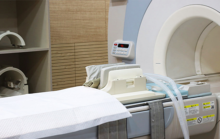
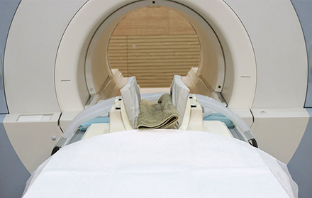
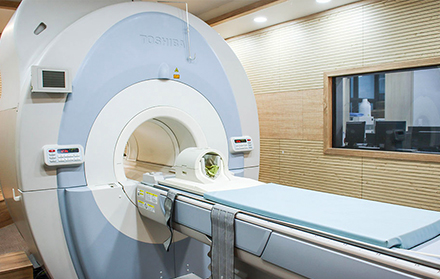
 1.5T high-field MRI
 FBI (Fresh Blood Imaging) reduces contrast agent use
 Shorter scan times and detailed images


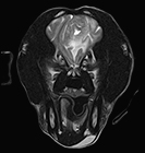 T2-weighted image
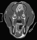 Post-contrast T1
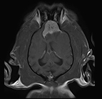 Post-contrast T1, coronal
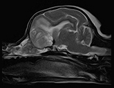 T2-weighted, parasagittal
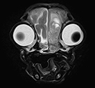 T2-weighted image
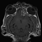 Post-contrast T1, coronal
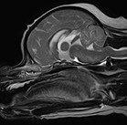 T2-weighted, sagittal
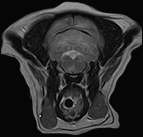 T2-weighted image
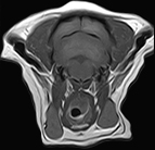 Post-contrast T1
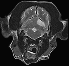 T2-weighted image
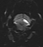 DWI
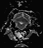 ADC map
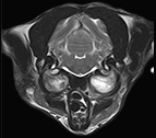 T2-weighted image
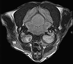 FLAIR
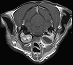 Post-contrast T1
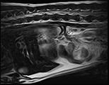 T2-weighted, sagittal
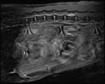 Post-contrast T1, sagittal
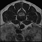 T2-weighted image
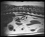 T2-weighted, sagittal
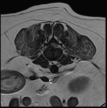 T2-weighted image
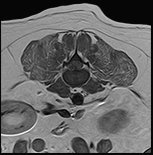 Post-contrast T1
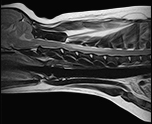 T2-weighted, sagittal
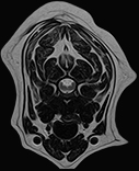 Post-contrast T1, sagittal
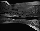 T2-weighted image

