


From patellar luxation, cruciate rupture and disc disease
to total joint replacement
Neuro & Orthopedic Surgery
-
01Arthroscopy
Arthroscopy
01Arthroscopy
Minimally invasive joint surgeryWhat is arthroscopy?
Like endoscopy, arthroscopy inserts an instrument with a miniature camera to look directly inside the joint — so diagnosis and treatment happen in the same procedure. Because the knee is not fully opened, an incision of only 0.5–1 cm allows accurate diagnosis and treatment, and the small incision helps reduce post-operative pain and shorten recovery.
Arthroscopy is performed by a veterinarian who completed arthroscopy courses in the Netherlands and at Colorado State University, with a focus on surgical safety and a fast recovery.
Benefits of arthroscopy
-

Minimally invasive
Surgery through a 0.5–1 cm incision instead of opening the whole knee joint. -

High accuracy
The arthroscope shows the affected area clearly while the surgeon operates. -

Less pain after surgery
A minimal incision usually means a faster recovery and less pain than conventional surgery. -

Diagnosis and treatment together
With the arthroscope, diagnosis and treatment can happen in a single procedure.
When we use arthroscopy
- Joint pain
- Lameness from a joint
- Joint inflammation
- Intra-articular fractures
- Ligament rupture
-
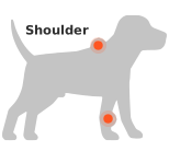 Removal of bone fragments from the shoulder and elbow
-
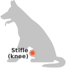 Confirming cruciate rupture and meniscal tear treatment
Arthroscopy course certificates
-
 Vetami arthroscopy course, Netherlands
-
 Colorado State University arthroscopy course
-
-
02Luxating Patella Surgery
Patellar Luxation Surgery
02Patellar Luxation Surgery
Kneecap dislocationWhat is patellar luxation surgery?
Patellar luxation is one of the most common orthopedic conditions, but cases differ. When it is complicated by cruciate ligament damage or by a deformed femur or tibia, the operation has to be tailored to the patient.
- 01Assess concurrent conditions Cruciate rupture, arthritis, limb alignment problems and other complicating factors
- 02Patient-specific surgical plan Based on the grade of luxation and the condition of the joint
- 03Anesthesia & sterile surgery Surgery with veterinarians trained in anesthesiology and a sterile operating system
- 04Post-operative recovery Rehabilitation and follow-up monitoring
Drawing on 10 years of clinical experience, patient-specific planning and a sterile system, we aim to reduce recurrence and after-effects. We also run a lifetime re-luxation policy with free revision surgery.
-
 Before surgery
-
 After surgery
-
 Re-luxation PolicyS Animal Medical Center
If the kneecap luxates again after surgery, we perform the revision surgery free of charge, for the life of your pet.
 Re-luxation policy certificate
-
03Cranial Cruciate Ligament Surgery
Cruciate Ligament Surgery
03Cruciate Ligament Surgery
Cranial cruciate ligament ruptureWhat is cruciate ligament surgery?
Our Surgery Center performs more than 100 TPLO operations a year. TPLO is widely regarded as the gold standard for cruciate ligament rupture. It is the most widely performed technique in countries with advanced veterinary care, with a low recurrence rate and a relatively fast recovery.
TPLO at S
- Accurate arthroscopic diagnosis · confirms the cruciate rupture and checks cartilage and other surrounding tissue for damage, through a minimal incision
- TPLO / CBLO for small to large dogs
- Nerve blocks for effective pain control and a faster recovery
After surgery, our rehabilitation program helps the joint and muscles recover.
-
 Before surgery
-
 After surgery
-
04Hip Surgery
Hip Surgery
04Hip Surgery
FHNO & total hip replacementWhat is hip surgery?
Hip surgery is performed when a hip problem makes walking difficult. The two main techniques are FHNO (femoral head and neck ostectomy) and THR (total hip replacement), most often for hip dysplasia and avascular necrosis of the femoral head (Legg-Calvé-Perthes disease). We choose the technique that suits the patient's weight, symptoms and disease stage.
Femoral head and neck ostectomy (FHNO)
FHNO removes the femoral head so it no longer contacts the acetabulum, allowing a fibrous false joint to form. It is a relatively simple, less costly procedure, but because the joint is not replaced, large dogs over 15–20 kg may have some restriction of movement afterward — so it is recommended mainly for small dogs.
Total hip replacement (THR)
THR replaces the damaged hip entirely with an artificial joint and can restore full joint function. It is a highly demanding operation, best performed by a dedicated orthopedic surgery team. At S Animal Medical Center we have:
- 01Surgery by a veterinarian who has completed THR certification training
- 02A dedicated, experienced THR surgical team
- 03Advanced THR workshop training
- 04Implant systems from BioMedtrix (large dogs) and Vetmaster (small dogs)
- 05Sterile operating rooms built to the standard for high-risk human operating rooms
Together with our experience in THR surgery, these help us reduce surgical risk and aim for a successful outcome.
-
 Before surgery
-
 After surgery
-
05Fractures and Shoulder Luxation
Fractures & Shoulder Luxation
05Fractures & Shoulder Luxation
Trauma surgeryFracture and shoulder luxation surgery
Fractures in dogs and cats from falls and traffic accidents vary in difficulty depending on their location and severity. We perform a range of fracture surgery: forelimb fractures, comminuted fractures (bone broken into several pieces), multiple fractures involving areas such as the pelvis and shoulder, and non-unions (bones that did not heal properly after surgery).
- 01CT-based 3D-printed surgical simulation The more complex the fracture, the more important it is to restore the exact angle and length of the bone. We print a 3D model from CT data, pre-contour the plates and rehearse the repair before surgery, to improve precision, shorten operating time and help make surgery safer.
- 02Minimally invasive fracture repair Interlocking nails and similar implants help limit damage to bone and surrounding muscle; arthrodesis (joint fusion) is used where the injury requires it.
- 03BMP (bone morphogenetic protein) Promotes bone formation and supports stable bone healing.
-
 Pre-operative 3D reconstruction and post-operative radiograph of a patient with multiple pelvic fractures after a traffic accident
-
 Minimally invasive repair with an interlocking nail
-
 Minimally invasive repair with an interlocking nail
-
 Radiographs before and after shoulder luxation corrected by arthrodesis
-
06Neurological Surgery
Neurosurgery
06Neurosurgery
Brain & spinal surgeryWhat is neurosurgery?
Neurological disease in dogs and cats can lead to nerve damage if treatment is delayed, so prompt diagnosis and treatment are important. With 1.5T MRI, detailed image reading by veterinarians trained in diagnostic imaging and a team-based surgical system, we diagnose and operate on disc disease, hydrocephalus, atlantoaxial instability (AAI), caudal occipital malformation syndrome (COMS, also called Chiari-like malformation), and brain and spinal cord tumors.
In neurosurgery we use the Sonopet ultrasonic aspirator to operate more precisely while limiting damage to nerve tissue, and after surgery a rehabilitation program — acupuncture, laser therapy and underwater treadmill — helps restore nerve function and the ability to walk.
Intervertebral disc herniation
A herniated disc presses on the spinal cord and can occur in the neck, back or lower back. In acute spinal cord injury, delay can mean permanent paralysis — so emergency surgery is sometimes needed.
After a neurological examination and MRI to establish an accurate diagnosis, we plan treatment — surgery, medication or non-surgical rehabilitation — and 24/7 Emergency Surgery at our Ulsan hospital helps emergency patients receive timely treatment.
Watch the video (Korean narration)COMS (Chiari-like malformation) · atlantoaxial instability (AAI) · complex lesions
Beyond COMS and AAI, which are common in small breeds, MRI-based localization helps us identify the actual cause of signs in complex cases where several lesions — disc disease, atlantoaxial (AA) band, syringomyelia — coexist, and plan treatment accordingly.
For AAI stabilization we build a CT-based 3D-printed model to analyze the anatomy before surgery and plan the exact fixation points.
-
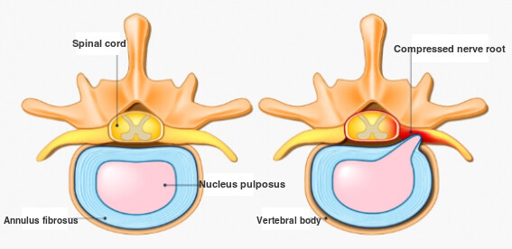 Signs of disc herniation
-
 Disc herniation surgery
-
 Radiograph of a patient who presented with seizures, was diagnosed with COMS on MRI, and underwent surgery
-


