


-
Eye screening
-
Cataracts
-
Glaucoma
-
Ocular surface disease
-
Corneal disease
-
Retinal disease
Comprehensive eye screening
For eye diseases that can lead to blindness, such as cataracts and glaucoma, early detection — and finding the right time for surgery — is very important.
Regular eye examinations are especially important for older pets.
Our eye screening program is called I Love Care; the tests it includes are listed below.
- History
- Basic eye examface, ocular symmetry, vision
- Tear production test
- Intraocular pressure
- Slit-lamp exameyelids, conjunctiva, third eyelid, cornea, anterior chamber, iris, front of lens
- Slit-lamp exam after dilationwhole lens, vitreous
- Fundus examretina, optic nerve, choroid
Depending on what the screening finds, we continue with:
- Discharge, redness or blinking
- Ocular surface work-up
- Microbiology
- Suspected corneal wound
- Corneal staining
- Anterior segment OCT
- Cataract — deciding on surgery
- Electroretinography (ERG)
- Ocular ultrasound
- Retina, optic nerve or vision abnormal
- Posterior segment OCT
- MRI
Screening items |
I Love Care | I Love Care+ | |
|---|---|---|---|
Screening items |
I Love Care | I Love Care+ | |
|---|---|---|---|
| History | We review vision, discharge, redness and other signs with you to assess your pet's current condition. | ||
| Neuro-ophthalmic exam | Assesses facial and ocular symmetry and basic function: vision, pupillary light reflex, palpebral reflex and more. | ||
| Tear production | Measures tear production on the eye surface. | ||
| Intraocular pressure | Measures the pressure inside the eye. | ||
| Slit-lamp exam (undilated) | Detailed examination of the structures around the eye — eyelids, conjunctiva, third eyelid — and inside it: cornea, anterior chamber, lens. | ||
| Slit-lamp exam (dilated) | After dilation, evaluates the back of the lens and the vitreous (essential for cataract assessment). | ||
| Fluorescein staining | Detects corneal wounds with fluorescein dye when injury is suspected. | ||
| Fundus exam | Checks the vitreous, retina and optic nerve for abnormalities affecting vision. | ||
| Chromatic PLR | Uses different wavelengths of light to detect retinal dysfunction. | ||
| Ocular ultrasound | Uses ultrasound to examine the inside of the eye, including when the slit lamp cannot give a clear view. | ||
| Gonioscopy | Examines the pectinate ligament, the front part of the aqueous outflow pathway. | ||
| UBM (ultrasound biomicroscopy) | Examines the ciliary cleft, the rear part of the aqueous outflow pathway. | ||
Cataracts
A cataract is a clouding of the lens that makes vision hazy. The further it progresses, the higher the risk of complications — and since surgery is currently the only treatment, having surgery at the right time improves the chance of success.
The cataract surgery process
-
01

Basic eye exam
Stage of the cataract and check for complications -
02

Pre-anesthetic tests
Screening for underlying disease and fitness for anesthesia -
03

Electroretinography
Tests retinal function to predict whether vision will return after surgery -
04

Cataract surgery
Choice of artificial lens and implants suited to the patient
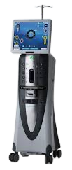
Cataract and retinal surgery
with the Stellaris Elite
The Bausch + Lomb Stellaris Elite allows precise control
and micro-incision surgery. It keeps intraocular pressure fluctuation within ±5 mmHg, which helps reduce the risk of retinal detachment and improves stability compared with earlier systems.
-
- Before
- After
-
- Before
- After
Glaucoma
Glaucoma damages optic nerve function through raised intraocular pressure or reduced blood flow to the nerve. As it progresses the risk of blindness rises and the damage cannot be reversed, so regular pressure checks matter. Glaucoma is an emergency: if you notice squinting, tearing, conjunctival redness or corneal swelling, please bring your pet in for an examination as soon as possible. If one eye already has glaucoma, protecting the other eye is important: appropriate preventive medication can help delay glaucoma in the second eye.
Examinations
- Slit-lamp examination
- Intraocular pressure
- Gonioscopy
- Optic nerve examination (OCT)
Glaucoma diagnosis and treatment pathway
- Intraocular pressure testElevated IOP — suspect glaucoma at 20 mmHg or above
- Eye drop therapyIf there is no response to drops:
- Vision present
- Ahmed valve implantation
- Laser cyclophotocoagulation
- No vision
- Intravitreal cidofovir injection
- Intraocular silicone prosthesis
- Enucleation
-
- Before
- After
Glaucoma valve implantation
-
- Before
- After
Intraocular prosthesis for glaucoma
Ocular surface disease
The ocular surface is made up of the cornea, conjunctiva, tear film, eyelids, lacrimal glands and meibomian glands. Injury, infection and other causes can lead to discomfort, light sensitivity and tearing — and to deteriorating vision. We offer a range of medical and surgical treatments to keep the ocular surface healthy.
Examinations
- Tear film
- Tear production
- Meibomian gland assessment
- Fluorescein and rose bengal staining
-
- Before
- After
Conjunctivitis treatment
-
- Before
- After
Keratitis treatment
Corneal disease
The cornea is the clear front surface of the eye that lets light in. Our goal is not only to preserve the shape of the eye but to keep the cornea as transparent as possible. Losing an eye because of a corneal injury is very rare, and outcomes are better when appropriate treatment starts early.
Examinations
- Slit lamp
- Optical coherence tomography (OCT)
-
- Before
- After
Corneal graft for corneal ulcer
-
- Before
- After
Conjunctival graft for corneal ulcer
Retinal disease
The retina lines the inside of the eye and sends visual information to the brain, so retinal disease calls for an integrated approach that considers the nervous system, not just the eye. Many retinal diseases are difficult to treat, but some can be — and a prompt, accurate diagnosis improves the chance of successful treatment.
Examinations
- Obstacle course (vision) test
- Fundus camera
- Ocular ultrasound
- Electroretinography (ERG)
-
- Normal
- Retinal detachment
Retinal detachment
-
- Normal
- Retinal atrophy
Retinal atrophy

